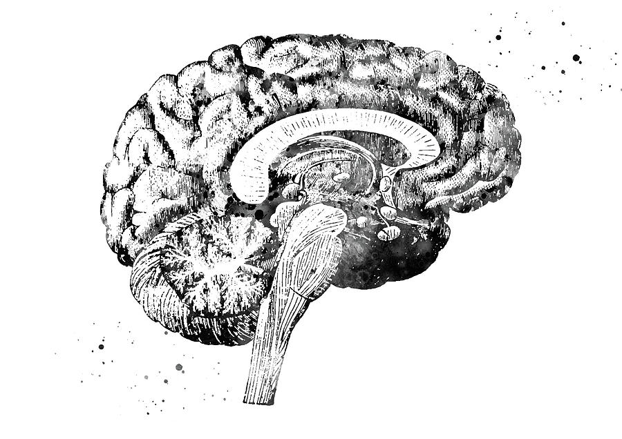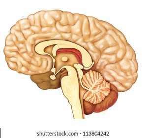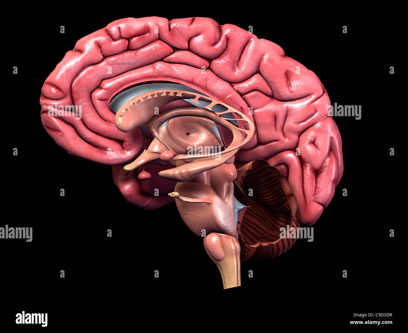

A few historical references are included to remind readers that many of the “discoveries” made with modern imaging techniques simply confirm what has been known about the temporal lobe for many years. References are provided for further reading, especially in areas where there is clinical interest or controversy or where anatomical details are not easily found in ordinary textbooks of neuroanatomy. Some physiological and pathological correlates are mentioned, but this is primarily an anatomical account. In this paper, I attempt to explain the positions of the parts of the normal human temporal lobe in relation to one another and to nearby structures. This article also reviews arterial supply, venous drainage, and anatomical relations of the temporal lobe to adjacent intracranial and tympanic structures. The temporal lobe contains much subcortical white matter, with such named bundles as the anterior commissure, arcuate fasciculus, inferior longitudinal fasciculus and uncinate fasciculus, and Meyer’s loop of the geniculocalcarine tract.

Its major projections are to the septal area and prefrontal cortex, mediating emotional responses to sensory stimuli. The amygdala receives input from the olfactory bulb and from association cortex for other modalities of sensation. The amygdala comprises several nuclei on the medial aspect of the temporal lobe, mostly anterior the hippocampus and indenting the tip of the temporal horn. The choroid fissure, alongside the fimbria, separates the temporal lobe from the optic tract, hypothalamus and midbrain. The largest efferent projection of the subiculum and hippocampus is through the fornix to the hypothalamus. Association fibres connect all parts of the cerebral cortex with the parahippocampal gyrus and subiculum, which in turn project to the dentate gyrus. The hippocampus is an inrolled gyrus that bulges into the temporal horn of the lateral ventricle. The hippocampal formation, on the medial side of the lobe, includes the parahippocampal gyrus, subiculum, hippocampus, dentate gyrus, and associated white matter, notably the fimbria, whose fibres continue into the fornix. All rights reserved.Only primates have temporal lobes, which are largest in man, accommodating 17% of the cerebral cortex and including areas with auditory, olfactory, vestibular, visual and linguistic functions. In conclusion, the MAPS and MAPS-HBSI methods give accurate and reliable volumes and atrophy rates across the clinical spectrum from healthy aging to AD.Ĭopyright 2010 Elsevier Inc. MAPS-HBSI required the lowest sample sizes (78 subjects) for a hypothetical trial. Comparing MCI subgroups (reverters, stable and converters): volumes were lower and rates higher in converters compared with stable and reverter groups (p< or =0.03). Both MAPS baseline volume and MAPS-HBSI atrophy rate distinguished between control, MCI and AD groups. The accuracy of our volumes was one of the highest reported (mean(SD) Jaccard Index 0.80(0.04) (N=30)). We compared our measures with those generated by Surgical Navigation Technologies (SNT) available from ADNI. The best method was applied to 682 ADNI subjects, at baseline and 12-month follow-up, enabling assessment of volumes and atrophy rates in control, mild cognitive impairment (MCI) and AD groups, and within MCI subgroups classified by subsequent clinical outcome.

Hippocampus anatomy cross section manual#
Methods were developed and validated against manual measures using subsets from Alzheimer's Disease Neuroimaging Initiative (ADNI). Change in volume over 12months (MAPS-HBSI) was measured by applying the boundary shift integral using MAPS regions.
Hippocampus anatomy cross section registration#
The improved technique uses non-linear registration of the best-matched templates from our manually segmented library to generate multiple segmentations and combines them using the simultaneous truth and performance level estimation (STAPLE) algorithm. We describe an improvement (multiple-atlas propagation and segmentation (MAPS)) to our template library-based segmentation technique. Delineation of the structure on MRI is time-consuming and therefore reliable automated methods are required. Volume and change in volume of the hippocampus are both important markers of Alzheimer's disease (AD).


 0 kommentar(er)
